Optical Coherence Tomography
Optical coherence tomography (OCT) is an imaging modality analogous to ultrasound, but instead of using the difference in the flight times of acoustic waves (as in ultrasound), it uses light to achieve micrometer axial resolution. OCT is used in many different biomedical applications, with retinal imaging being the most successful and the driving force behind much OCT development. The axial resolution of OCT in retinal tissue is about 1-15 µm, which is 10 to 100 times better than ultrasound or MRI. Although relatively new to ophthalmology, a commercial OCT system has already revolutionized the field, rapidly becoming an essential tool in the diagnosis and monitoring of human retinal disease. As part of our collaboration with the Biomedical Engineering Department at Duke University, our group at the Vision Science and Advanced Retinal Imaging Laboratory has developed a Fourier-domain OCT system that is faster and has higher resolution than existing commercial OCT instruments. In collaboration with colleagues in the Computer Science Department at UC Davis, we have also developed software for three-dimensional rendering to allow visualization and quantitative evaluation of volumetric data acquired with Fourier-domain OCT.
Background
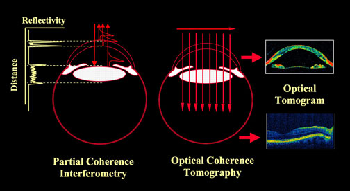
The principle of ocular imaging with OCT is based upon measuring the time-delay of the light reflected from each optical interface (A-scan) when a pencil of light enters the eye. A series of A-scans across the structure permits a cross-sectional reconstruction of a plane through the anterior or posterior segment of the eye. This is known as a B-scan. Because the velocity of light is so high, and the distance between layers is so short (µm), it is not possible to measure the flight-time change directly. Instead, OCT uses low coherence interferometry in which the light source is split between that entering the eye and a reference path. The light reflected back from the two paths forms an interference pattern when the distance in the two paths matches to within the coherence length of the light source. Due to the coherent detection (multiplication of the reference and sample arm), OCT allows measurements of weakly reflecting retinal layers (on the order 10-6 to 10-9). This may be measured in the time domain (taking into account the position of a reference mirror or optical delay line in the reference channel), or in the Fourier-domain using a spectrometer or sweeping the light source wavelength and calculating the inverse Fourier transform as a function of wavelength. OCT systems may have a sensitivity of ~100 dB over a range of approximately 2-3 mm. Because of the high resolution of OCT, it is possible to obtain images in the living eye that previously were only possible by removing the tissue and examining it under the microscope.

OCT at UC Davis
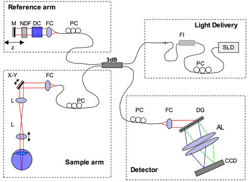
A number of different types of OCT imaging have been developed for the human eye including time-domain and Fourier-domain techniques. The most popular time-domain system configurations include standard A-scan acquisition, transverse scanning and full-field OCT. Among Fourier-domain methods, spectral and swept-source OCT are most commonly used. Over the years several variations of functional OCT have been developed for both time-domain and Fourier-domain systems, including Doppler (flow), polarization-sensitive and spectroscopic OCT. The OCT system constructed in our laboratory uses the Fourier-domain (spectrometer based) approach. The main advantages of Fourier-domain OCT are higher signal acquisition rates and higher sensitivity than time-domain OCT. The figure below shows a schematic of our system at UC Davis.

The light source is a superluminescent laser diode (SLD) that provides up to 3.5 µm axial resolution in the retina. The light is passed through a polarization controller (PC) and Faraday isolator (FI) before being split between the reference and sample arms. The light in the reference arm must pass through a water vial to compensate for the dispersion (DC) of the eye, before reaching the reference mirror (M). The light in the sample channel is scanned across the retina under computer control. The reference and sample channels are combined and the light passes through a transmissive diffraction grating (DG) and is imaged onto a CCD camera.
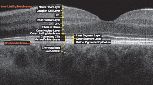
Here we show a cross-sectional, or B-scan, of a normal human retina with the layers identified. OCT depends upon different optical properties than microscopy so the proper labeling of retinal layers is still being studied.

Because Fourier-domain OCT is fast, it is possible to acquire multiple B-scans, register (align) them and construct a three-dimensional view of regions of interest. One can “slice” the image in any orientation such as through the top of the retina to obtain C-scans, which provide the view that is similar to that obtained with fundus cameras or scanning laser ophthalmoscopes. The movie below shows the three-dimensional structure of the optic nerve head of a normal individual and successive C-scans that penetrate the optic nerve head into the nerve itself.
Double-click to view movie
Even the lower resolution OCT systems are becoming an indispensable tool for practicing ophthalmologists. With higher resolution systems new findings are emerging almost daily. Fourier-domain OCT may be used not only for diagnosis and monitoring of retinal disease, but is an especially valuable tool for research. Here we show but one example from our laboratory, a patient diagnosed with central serous chorioretinopathy (fluid build up causing retinal layers to become detached).
Double-click to view movie
