Reading Center
The UC Davis Eye Center hosts the Visual Field and OCT Reading Center to support clinical research. John L. Keltner and John S. Werner are the co-directors. The primary aim of the Reading Center is to ensure, through rigorous quality control, the validity and integrity of visual field and OCT data. The Visual Field Reading Center (VFRC) routinely conducts quality control assessment of visual field data, provides expertise in the collection and inspection of visual field data, and provides training, certification and continuous feedback regarding technician performance. To date, approximately 77,000 visual fields (7,256 qualifying assessment and 69,630 follow-up fields) have been received, processed and monitored by the Center.

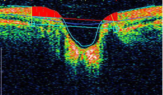
Stratus OCT image with Segmented RNFL in Glaucoma
The VFRC has served as the reading center for the Ocular Hypertension Treatment Study (OHTS) since 1994. This was the largest glaucoma clinical trial in the United States. To date, only 3.6% (2,540/69,630) of OHTS follow-up fields are unreliable, thus confirming the fact that strict quality control assessment procedures provide more reliable and valid visual field data. Since 1988, the VFRC has also been the reading center for the 15-year follow up to the Optic Neuritis Treatment Trial and has evaluated approximately 18,000 visual fields.

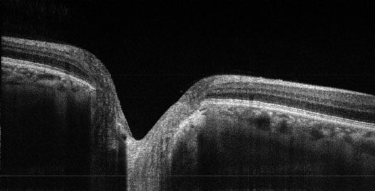
High-Resolution Image of Optic Nerve Head
(UC Davis OCT instrumentation)
We are building on the framework established for quality control of visual fields in clinical trials and our state-of-the-art OCT development to provide an OCT reading center. Our OCT development is supported by funding for a Bioengineering Research Partnership from the National Eye Institute (John S. Werner, Principal Investigator). This center processes images from OCT3 (Zeiss Meditec) using the manufacturer’s software as well as programs developed at UC Davis for image segmentation. The importance of an OCT reading center for clinical trials is highlighted by recent publications indicating that overall retinal thickness and nerve fiber layer thickness using standard algorithms are incorrect a high percentage of time. Reliability of experienced readers and custom segmentation software provides quality control assessment needed for clinical trials. Using our custom software, it is possible to obtain precise measurements of structures that are not included in Stratus software.

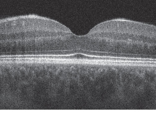
High-Resolution Image of Fovea
(UC Davis OCT instrumentation)
Our group also has extensive experience in measurement of contrast sensitivity and visual acuity using custom and standardized devices. We are prepared to assist in the calibration of devices used in clinical trials and to provide quality control across testing sites for studies involving measurement of contrast sensitivity and acuity.

Contrast Sensitivity (contrast vs. spatial frequency)
For further information, please contact Dr. John L. Keltner at the UC Davis Medical Center (jlkeltner@ucdavis.edu).
RNFL Segmented in Three Dimensions using Custom Software
