Publications
Recent highlights
For a full list of publications go to Google Scholar, ORCID, or PubMed.
The past few years have witnessed rapid development of the optoretinogram—a noninvasive, optical measurement of neural function in the retina, and especially the photoreceptors (Ph). While its recent development has been rapid, it represents the culmination of hundreds of experiments spanning decade…
Jonnal, Ravi S.
Full-field swept-source optical coherence tomography (FF-SS-OCT) is an emerging technology with potential applications in ophthalmic imaging, microscopy, metrology, and other domains. Here we demonstrate a novel method of multiplexing FF-SS-OCT signals using carrier modulation (CM). The principle of…
Valente, Denise; Vienola, Kari V.; Zawadzki, Robert J.; Jonnal, Ravi S.
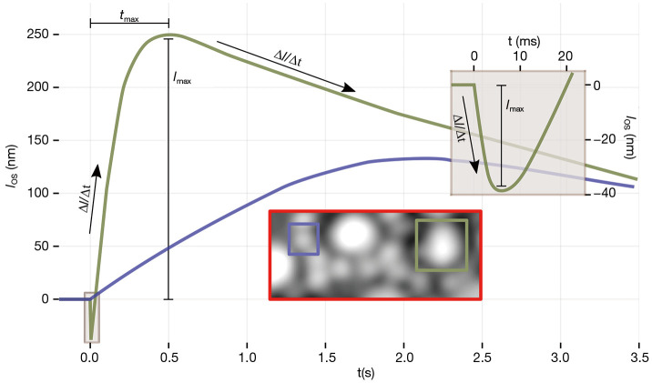
Noninvasive, objective measurement of rod function is as significant as that of cone function, and for retinal diseases such as retinitis pigmentosa and age-related macular degeneration, rod function may be a more sensitive biomarker of disease progression and efficacy of treatment than cone functio…
Azimipour, Mehdi; Valente, Denise; Vienola, Kari V.; Werner, John S.; Zawadzki, Robert J.; Zawadzki, Robert J.; Jonnal, Ravi S.
A retinal imaging system was designed for full-field (FF) swept-source (SS) optical coherence tomography (OCT) with cellular resolution. The system incorporates a real-time adaptive optics (AO) subsystem and a very high-speed CMOS sensor, and is capable of acquiring volumetric images of the retina a…
Valente, Denise; Vienola, Kari V.; Zawadzki, Robert J.; Zawadzki, Robert J.; Jonnal, Ravi S.
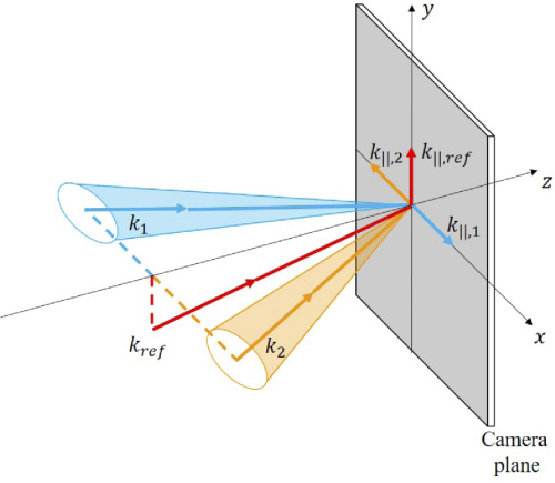
We describe the details of a multimodal retinal imaging system which combines adaptive optics (AO) with an integrated scanning light ophthalmoscopy (SLO) and optical coherence tomography (OCT) imaging system. The OCT subsystem consisted of a swept-source, Fourier-domain mode-locked (FDML) laser, wit…
Azimipour, Mehdi; Jonnal, Ravi S; Werner, John S; Zawadzki, Robert J
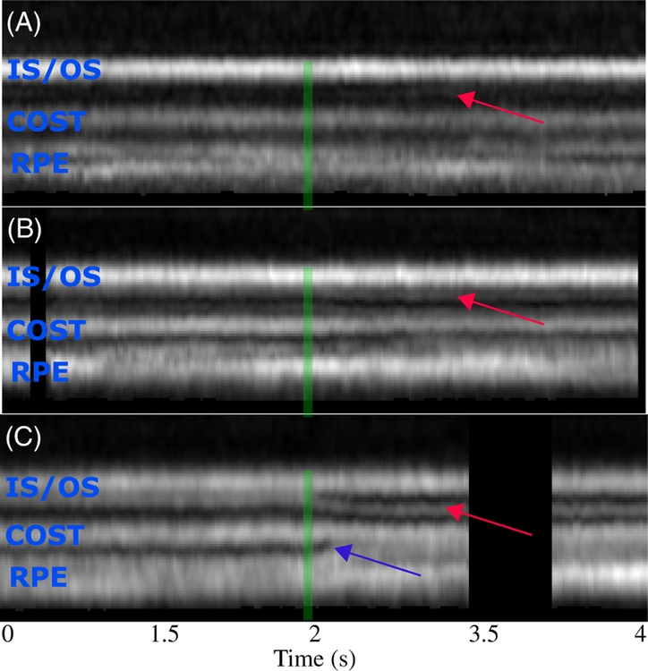
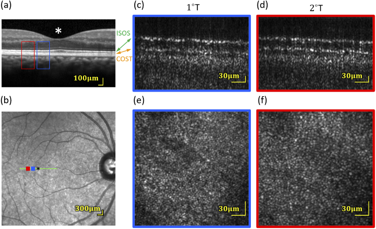
Objective optical assessment of photoreceptor function may permit earlier diagnosis of retinal disease than current methods such as perimetry, electrophysiology, and clinical imaging. In this work, we describe an adaptive optics (AO) optical coherence tomography (OCT) system designed to measure func…
Azimipour, Mehdi; Migacz, Justin V; Zawadzki, Robert J; Werner, John S; Jonnal, Ravi S

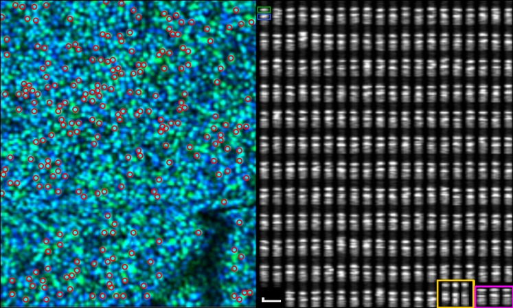
In retinal raster imaging modalities, fixational eye movements manifest as image warp, where the relative positions of the beam and retina change during the acquisition of single frames. To remove warp artifacts, strip-based registration methods-in which fast-axis strips from target images are regis…
Azimipour, Mehdi; Zawadzki, Robert J; Gorczynska, Iwona; Migacz, Justin; Werner, John S; Jonnal, Ravi S
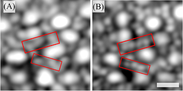
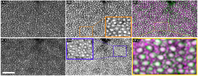
Purpose: Optical coherence tomography’s (OCT) third outer retinal band has been attributed to the zone of interdigitation between RPE cells and cone outer segments. The purpose of this paper is to investigate the structure of this band with adaptive optics (AO)-OCT. Methods: Using AO-OCT, images wer…
Jonnal, Ravi S; Gorczynska, Iwona; Migacz, Justin V; Azimipour, Mehdi; Zawadzki, Robert J; Werner, John S
Patents
Miller, Donald T., Ravi S. Jonnal, Junle Qu, and Karen E. Thorn. “Method and apparatus for improving both lateral and axial resolution in ophthalmoscopy.” U.S. Patent 7,364,296, issued April 29, 2008.